How plant based diets can help people with rheumatoid arthritis

Many recent studies have highlighted the health benefits that a plant based diet can bring. Now, a new review explains just why it can be helpful for people living with rheumatoid arthritis.

Rheumatoid arthritis — a chronic autoimmune condition that causes pain and stiffness in the joints — has a prevalence of between 0.3% and 1% among the world’s population, according to the World Health Organization (WHO).

The condition can become so debilitating as to stop people from continuing in full time work. As the WHO also note, within only 10 years from disease onset, at least 50% of individuals with rheumatoid arthritis in high income countries become “unable to hold down a full time job.”

Doctors usually prescribe a range of drugs and lifestyle adjustments to help people manage their rheumatoid arthritis and make disability less likely. Management strategies that healthcare providers might advise include increased physical activity and weight loss.

Now, a new review appearing in the journal Frontiers in Nutrition shows that following a plant based diet can be a useful intervention when it comes to coping with this condition, as it triggers some helpful biological changes.

‘Symptoms may improve or even disappear’

The review — conducted by specialists from the Physicians Committee for Responsible Medicine in Washington, DC — looked at recent studies that observed the impact of diet on biological mechanisms that are important in rheumatoid arthritis.

It concluded that plant based diets lead to specific changes that can help relieve the symptoms of this condition.

One key way in which plant based diets can be helpful is by reducing levels of inflammation. The review authors cite a study from 2015 that showed participants who ate a plant based diet for 2 months had lower inflammation than those who ate a diet that was high in fat and featured more animal products.

The team also notes that additional research has found an association between adherence to diets high in fat and processed meat and a rise in markers of inflammation. One of these markers is C-reactive protein, a protein present in the blood, and one which reacts to inflammation.

On the other hand, following plant based diets or diets that have a high content of fiber has an association with lower levels of C-reactive protein.

Another study that the review looked at was a randomized clinical trial showing that, after following a low fat vegan diet for 4 weeks, individuals with moderate-to-severe rheumatoid arthritis saw significant improvements in symptoms, including joint pain and stiffness, tenderness, and swelling.

People with rheumatoid arthritis can also benefit from losing extra weight. According to evidence from a 2018 study, overweight individuals with rheumatoid arthritis who shed in excess of 5 kilograms were three times more likely to have improvements in their symptoms compared to those who lost less weight.

The review authors explain that vegetarian and vegan diets appear to help people lose weight, more so than any other diet types.

Finally, the researchers explain that plant based diets also seem to promote a healthy gut environment, since many of these diets are high in fiber, which, as studies have shown, influences the composition of the gut microbiome.

Specifically, plant based diets seem to increase bacterial diversity in the gut, which could help people with rheumatoid arthritis, precisely because they tend to lack bacterial diversity.

The investigators who conducted the review suggest there is a need for further research into the benefits that plant based diets may afford to people with inflammatory autoimmune conditions, as well as their underlying mechanisms.

However, they note that, so far, the emerging evidence suggests that eating more fruits, vegetables, whole grains, and legumes could make a real difference for people with rheumatoid arthritis.

“A plant based diet comprised of fruits, vegetables, grains, and legumes may be tremendously helpful for those with rheumatoid arthritis. This study offers hope that with a simple menu change, joint pain, swelling, and other painful symptoms may improve or even disappear.”

Study co-author Dr. Hana Kahleova

A complex relationship between Alcohol & Dementia

Although alcohol has been popular for millennia, and dementia is increasingly prevalent, scientists are yet to understand the relationship between the two. A recent study sets out for answers.

With dementia predicted to affect 13.9 million adults in the United States by 2060, understanding why these conditions develop is more urgent than ever.

Scientists have uncovered certain factors that increase the risk of developing dementia. Some, such as advancing age, cannot be prevented. However, it is possible to avoid other potential risk factors, such as smoking tobacco.

It is essential to identify modifiable risk factors as understanding these could help prevent or delay the onset of dementia.

Recently, researchers designed a study to look for links between dementia and alcohol consumption in older adults. They published their findings in JAMA Network Open.

What do we already know?

Perhaps surprisingly, as the authors explain, “we know little about the independent associations of quantity and frequency of alcohol consumption with dementia risk.”

Although some studies have investigated alcohol and dementia more broadly, there are still substantial gaps in our understanding. For instance, in an earlier study, researchers calculated alcohol consumption as a daily average across each year.

Using a daily average in this way misses the nuances of alcohol quantity and frequency.

This is an important point. For example, drinking 7 glasses of beer on 1 day each week is likely to have a different impact than drinking 1 glass of beer each night, every day of the week; although the quantity consumed is the same.

The authors of another study concluded that binge drinking in midlife increases the risk of dementia. However, it is still not clear whether regularly drinking smaller amounts of alcohol has the same effect.

Other studies muddy the water further by looking at the links between alcohol, dementia risk, and the presence of apolipoprotein E4 (APOE E4). This gene variant has associations with an increased risk of developing Alzheimer’s.

An earlier study concluded that alcohol consumption increases the risk of dementia and that people with the APOE E4 variant have a higher chance of developing dementia.

Another unknown is how alcohol influences dementia risk for individuals with mild cognitive impairment (MCI). Scientists consider MCI to be the stage between normal age-related cognitive decline and dementia.

Are you looking for alternative health insurance?

Take a short assessment and find affordable short-term health insurance, indemnity insurance, and supplemental health products — like pharmacy benefits and emergency medical plans — to fit your needs.

Alcohol and dementia data

To investigate, the scientists delved into an existing dataset, extracting the information from 3,021 participants, with an average age of 72.

The Ginkgo Evaluation of Memory Study from 2000–2008 supplied the data.

At the beginning of the study, participants provided information about the amount of alcohol they consumed, how often they drank, and what types of drinks they consumed — beer, wine, or liquor.

According to the authors, at the start of the study, each participant went through “a comprehensive neuropsychological battery of 10 tests.” These assessed a range of cognitive functions, and participants completed the tests every 6 months.

Scientists isolated DNA from blood samples to identify carriers of the APOE E4 variant.

Alongside genetic data and details about alcohol use, the scientists also collated information about blood pressure, body weight, height, history of heart disease, and smoking status. They also asked questions to estimate how regularly each participant was involved in social interactions.

Many gaps remain

Of the 3,021 participants, 2,548 did not have MCI, and 473 did have MCI. Over approximately 6 years of follow-up, 512 participants received a diagnosis of dementia.

The authors found that, among those without MCI, no amount of alcohol consumption increased dementia risk compared with those who drank less than one drink per week.

When they analyzed the group of individuals with MCI, it was a similar story; there was no statistically significant difference.

However, dementia risk appeared to be highest for the individuals with MCI who drank 14 drinks per week compared with those who drank less than one drink each week — a relative risk increase of 72%.

The authors also note some differences associated with drinking patterns:

“Daily low-quantity drinking was associated with lower dementia risk than infrequent higher-quantity drinking among participants without MCI at baseline.”

Although the authors are keen to underline the negative impacts of excessive drinking on brain health and health in general, they also note that:

“Our findings provide some reassurance that alcohol consumed within recommended limits was not associated with an increased risk of dementia among older adults with normal baseline cognition.”

When the researchers investigated the potential impact of APOE E4, they found no significant effect, which mirrors several previous studies. The authors theorize that the lack of effect might be because the association is more pronounced in younger populations; they call for further investigation.

In conclusion, the current study provides few solid answers. It confirms, however, that the relationship between alcohol and dementia is complex and likely to require a great deal more research.

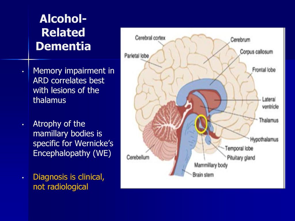
Memory impairment in ARD correlates best with lesions of the thalamus. Atrophy of the mamillary bodies is specific for Wernicke’s Encephalopathy (WE) Diagnosis is clinical, not radiological.

Link found between chronic headache and back pain

A review of 14 studies found that people with persistent headache or back pain were twice as likely to experience the other disorder as well.

Chronic headaches and persistent back pain are both debilitating conditions. New findings suggest a link between the two, potentially charting a new course for more effective treatment.

Chronic headaches and back pain both appear in the top five causes of years lived with disability. Healthcare professionals often treat the conditions separately, but there is a theory that in some people, they appear together. Therefore, treating both as one disorder may provide better results.

Up to 4% of individuals in the global adult population have headaches on 15 or more days of every month, according to the World Health Organization (WHO).

Meanwhile, about 80% of adults experience low back pain at least once in their lifetime, according to the National Institute of Neurological Disorders and Stroke, and approximately 20% of these people go on to develop chronic low back pain.

In 2013, a German study found a link between low back pain and both chronic migraine and chronic tension-type headaches.

Now, researchers from the University of Warwick, United Kingdom, appear to have found an even stronger association.

Double the odds

Appearing in the Journal of Headache and Pain, their review involved 14 studies with a range of sample sizes. The smallest study included 88 participants, while the largest involved 404,206 individuals.

One type of headache and a particular back pain disorder were the focus: chronic headaches and persistent low back pain. The team notes the definitions of these conditions, describing the former as headaches that occur on the majority of days for a minimum of 3 months and the latter as pain “between the bottom of the rib cage and the buttock creases” for the same period.

The review found that all the studies shared a similar positive association between the two conditions. However, the odds of experiencing both conditions varied widely between the studies from less than twice as likely to eight times as likely.

This meant that the researchers were unable to pool the data in a combined statistical analysis. However, Prof. Martin Underwood notes, “[i]n most of the studies, we found that the odds were about double — either way, you’re about twice as likely to have headaches or chronic low back pain in the presence of the other.”

For people experiencing migraine ⁠— the third most prevalent illness in the world ⁠— the link was even stronger.

A few limitations exist, namely that inconsistent study designs and populations and varying definitions of the disorders may have weakened the quality of the results.

Still, the findings are “very interesting,” notes Prof. Martin Underwood, from Warwick Medical School, “because typically these have been looked at as separate disorders and then managed by different people.”

“But this makes you think that there might be, at least for some people, some commonality in what is causing the problem.”

Combining treatments

The researchers are not yet sure of the potential shared cause, but they have some theories. One, explains Prof. Underwood, is that there may be “an underpinning biological relationship” in some people with headaches and back pain.

“There may be something in the relationship between how people react to the pain,” he says, “making some people more sensitive to both the physical causes of the headache, particularly migraine, and the physical causes in the back, and how the body reacts to that and how you become disabled by it.”

Such a cause could be a target for treatment. In fact, the team thinks that a joint approach may be best.

Right now, drugs are available to treat chronic headaches. Some people try a range of holistic remedies too. Back pain, meanwhile, may require a combination of exercise and psychological therapy.

Ensuring that people with both disorders receive appropriate support and management techniques could well help their quality of life. A combined treatment method could also provide a financial benefit by reducing the number of medication prescriptions.

“There is a need for doctors and other healthcare professionals to think that when treating one issue, [they should] ask about the other and tailor the treatment accordingly.”

Prof. Martin Underwood

“For future research, there’s probably work that needs to be done to understand what the underlying mechanisms behind this relationship are,” he adds.

Man having a back pain over gray background

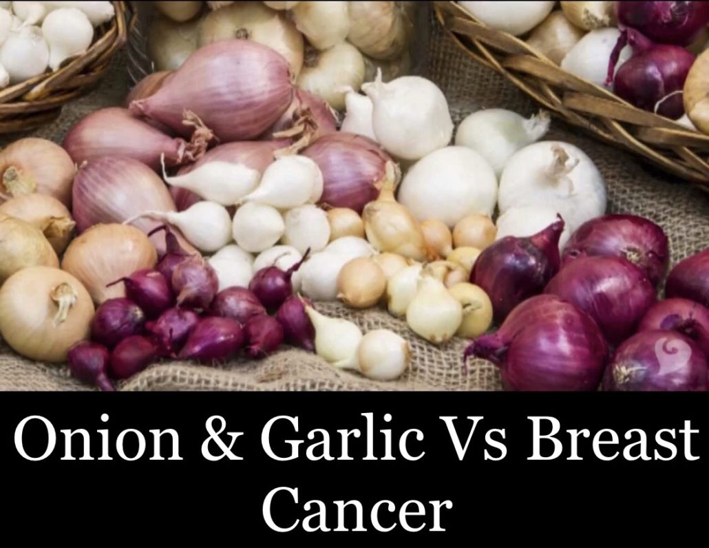
Onion & Garlic Vs Greasy Cancer

Onions and garlic may protect against breast cancer

New research examines the consumption of onion and garlic among women in Puerto Rico and suggests that the vegetables can significantly reduce the risk of breast cancer.

Onions and garlic are part of the allium genus, along with leeks, chives, and hundreds of other species.

Rich in flavor, with a strong, pungent kick, these “feisty” vegetables may also be able to fight off diseases, such as diabetes, heart disease, and cancer, according to emerging evidence.

In regards to cancer, several studies have specifically explored the links between onion and garlic consumption and the risk of colorectal, stomach, and prostate cancers.

Overall, these studies have found that the more of these vegetables that people consume, the lower their risk of developing these cancers.

However, less extensive research has gone into the link with breast cancer, say researchers led by Gauri Desai, a doctoral candidate in epidemiology at the University at Buffalo (UB), part of The State University of New York.

So, Desai and the team set out to investigate this topic further, looking at a population of women in Puerto Rico. The reasons for choosing this population were twofold. Firstly, “Puerto Rico has lower breast cancer rates, compared to the mainland [United States], which makes it an important population to study,” explains Desai.

Secondly, a condiment called “sofrito,” which consists largely of garlic and onion, is a traditional Puerto Rican sauce that is widely consumed across the island.

The researchers published the results of their findings in the journal Nutrition and Cancer.

Studying onions, garlic, and cancer risk

Desai and the team used hospital and clinic records to identify 314 women aged between 30 and 79 who had breast cancer between 2008 and 2014. The researchers also included 346 control participants who were matched based on age and residential area.

nonmelanoma skin cancer. The researchers used a food frequency questionnaire to enquire about dietary patterns and total onion and garlic intake, which included sofrito consumption.

The team employed unconditional logistic regression to examine the association between onion and garlic intake and breast cancer occurrence, adjusting for factors such as age, education, family history, body mass index, smoking status, and others.

Sofrito linked to 67% lower breast cancer risk

The researchers found an inverse association between moderate and high total onion and garlic consumption and breast cancer cases, compared with low consumption of these vegetables.

Their findings were consistent after the researchers had stratified the results according to menopausal status, suggesting that “High onion and garlic consumption is protective against breast cancer in [the studied] population.”

Specifically, the authors write, “Sofrito intake, when examined alone, was inversely associated with breast cancer; for those consuming sofrito more than once/day, there was a 67% decrease in risk, compared to never consumers.”

“We found that among Puerto Rican women, the combined intake of onion and garlic, as well as sofrito, was associated with a reduced risk of breast cancer.”

Gauri Desai

“Studying Puerto Rican women who consume a lot of onions and garlic as sofrito was unique,” Desai adds, referring to one strength of the research.

Why might onions, garlic lower cancer risk?

Study co-author Jo Freudenheim, Ph.D., chair of epidemiology and environmental health at UB, also comments on the uniqueness of the study, saying, “There is very little research on breast cancer in Puerto Rico.”

“This study was a collaboration between my colleagues here at UB and at the University of Puerto Rico to help us understand why rates there are lower than in the rest of the U.S., and why rates there are continuing to increase, while they are decreasing in the rest of the [U.S.].”

Although the study was observational and cannot explain the mechanisms behind the findings, the researchers suspect that the flavonols and organosulfur compounds abundant in onions and garlic may be responsible for the anticancer effects.

In particular, the S-allylcysteine, diallyl disulfide, and diallyl sulfide in garlic and the alk(en)yl cysteine sulphoxides in onions have shown “anticarcinogenic properties in humans, as well as in experimental animal studies,” says Dr. Lina Mu, an associate professor of epidemiology and environmental health at UB and the study’s senior author.

Some study limitations

However, the authors also recognize some limitations to their research. For instance, they write, “The size of the study was small, particularly for analyses stratified on menopausal status.”

Secondly, the number of people who had never consumed garlic or onion was too small to use as a reference group. Therefore, the low exposure group still consumed some onion and garlic.

For this reason, say the researchers, the results “may have underestimated the true association between garlic and onion intake and breast cancer.”

“Furthermore, there is not a standardized recipe for sofrito, a condiment which is frequently homemade and therefore varies at least somewhat in recipe from person to person,” add the authors. “Hence, we were also not able to estimate the amount of onion and garlic in sofrito.”

“Although the recipe for sofrito varies to some extent, other ingredients, such as bell peppers, tomatoes, cilantro, and black pepper are usually added. Since we did not adjust our models for these ingredients, we cannot be sure that our results were due to the sole effect of onions and garlic.”

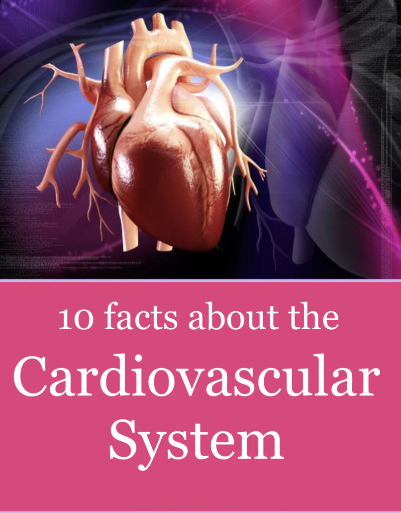
KNOW YOUR HEART. 10 Facts about the Cardiovascular system…

The cardiovascular system carries blood and nutrients to the cells of the body. The function of this system has effects on other parts of the body. A nursing student has many facts to learn about the cardiovascular system. Therefore, this article below will start you off with a list of 10 facts about the cardiovascular system that every nursing student should know.

The cardiovascular system or circulatory system consists of the heart, blood and blood vessels. The heart is the pump of the system and sits in the thoracic cavity. The heart sits predominately on the left side. Therefore, approximately two-thirds of the heart is to the left side of the sternum. The blood is a connective tissue. It is the fluid component of the system. The blood is transported to the cells via a network of passageways call the blood vessels.

There are 3 major layers of the heart. The layers are the endocardium, myocardium and the pericardium. The endocardium lines the inner chambers of the heart and the valves. The myocardium makes up the heart wall. The pericardium is the container of the heart. This triple-walled layer protects the heart. Also, the pericardium contains a fibrous layer and a serous layer. The serous layer has two separate layers. These layers are the parietal and visceral layers.

Below is a list of 10 facts about the cardiovascular system that every nursing student should know to help build a foundation of knowledge of the cardiovascular system.

Fact #1: The Cardiovascular System Is A Closed System.

The cardiovascular system is a closed system and also a system which is under pressure. This means if there is a leak in a large vessel it does not drip it sprays, especially a leak on the arterial side.

Therefore, when small leaks occur the system has a method of stopping these leaks. Hemostasis is the system within the blood that stops these leaks. Hemostasis is defined as the stoppage of blood. This system is constantly in action sealing small insults to the system.

Substances contained in the blood assist the process of hemostasis. The blood contains calcium ions and plasma proteins that cause coagulation or clotting within seconds of an injury. These plasma proteins are your clotting factors. (e.g. prothrombin and vitamin K)

Fact #2: The Heart Has Four Chambers.

The heart has four chambers: the right and left atria and the right and left ventricles. The right atrium and right ventricle receive deoxygenated blood from the body. The left atrium and left ventricle receive oxygenated blood from the lungs.

The atria of the heart are mostly reservoirs. The atria only contribute “atrial kick” to the cardiac cycle. Atrial kick or atrial contraction contributes approximately 20% of the volume to ventricular filling.

The ventricles make up the majority of the heart. The ventricles of the heart receive blood from the atrium. They eject blood into the pulmonary system (lungs) and to the systemic circulation (body).

The right side of the heart (ventricle) pumps against a low-pressure system (pulmonary circulation) and the left side of the heart (ventricle) pumps against a high-pressure system (systemic circulation). The left ventricle works harder than all the other chambers because it has to pump against the high pressure of the systemic circulation.

Fact #3: The Heart Has Four Valves.

The right side of the heart contains the tricuspid and pulmonary valves. The left side of the heart contains the bicuspid (mitral) and aortic valves.

The tricuspid and bicuspid (mitral) valves separate the atrium and ventricles.  These valves are also called the atrioventricular or AV valves.  As you would guess the tricuspid valve had three cusps or leaflets and the bicuspid valve has two cusps or leaflets.

The pulmonary valve opens to the pulmonary circulation and the lungs. The aortic valve opens to the systemic circulation. These valves are also called the semilunar valves because of their shape. The pulmonary and aortic valve each has three cusps or leaflets that are shaped like a half moon.

The “Lub-Dub” sound you hear with your stethoscope is the closing of the heart valves. The heart sound S1 is the closure of the tricuspid and bicuspid (mitral) valves. The heart sound S2 is the closure of the pulmonary and aortic valves.

Fact #4: The Heart Valves Operate Due To A Pressure System.

The valves of the heart open and close due to pressure within the system. During the cardiac cycle, the atria fill with blood. As the atria fill, the pressure in the atria eventually exceeds the pressure in the ventricles. When this happens the tricuspid and bicuspid (mitral) valves open and blood flows into the ventricles.

As blood flows into the ventricles the pressure begins to rise. Eventually, the pressure in the ventricles exceeds the pressure in the atria. This pressure that builds up in the ventricles is attributed to filling volumes of the ventricles. At this time the pulmonary valve and aortic valves close.

Following the isovolumetric contraction of the ventricles, the pulmonary and aortic valves open. Then, blood is ejected into the pulmonary circulation and systemic circulation.

Both the left and right atria fill at the same time and both the left and right ventricles fill at the same time.

Fact #5: Blood Vessels Are The Vascular Portion Of The Cardiovascular System.

Blood vessels include arteries and veins. When the blood leaves the heart it flows into the arteries. Arteries carry oxygenated blood from the heart to systemic circulation. The arteries divide into the smaller arterioles. Next, the arterioles divide into the even smaller capillary network on the arterial side. This arterial capillary network feeds the cells.

The veins divide into smaller venules. The venules divide into the smaller capillary network of the veins. Starting at the capillary network, the capillaries on the vein side pick up carbon dioxide and waste products which travel to the venules and then to the veins and back to the heart.

Blood flows from the heart to the arteries to the arterioles to the arterial capillary network. Then blood moves from the vein capillary network to the venules to the veins and back to the heart.

Fact #6: Blood Vessels Can Constrict And Dilate Having An Effect On Blood Pressure.

The sympathetic nervous system controls the blood vessels. Blood vessels have the ability to constrict or dilate with signals from the sympathetic nervous system.

Vasoconstriction and vasodilation occur when the blood vessels dilate and constrict. Vasoconstriction causes a decrease in the inner diameter of the blood vessel. Vasodilation causes an increase in the inner diameter of the blood vessel.

Blood vessels have an effect on blood pressure.

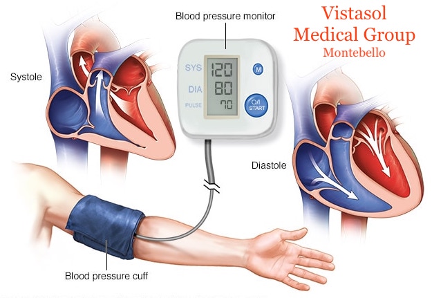
Remember, blood pressure is the measure of the pressure exerted on the walls of the blood vessel. The greater the pressure within the blood vessel the higher the blood pressure measurement. The lower the pressure within the blood vessel the lower the blood pressure measurement. The systolic pressure is the maximum pressure against the wall of the blood vessel and the diastolic pressure is the recoil.

A change in the diameter of the blood vessels causes changes in the blood pressure. When the blood vessels constrict (vasoconstriction), the blood pressure is higher. This is because the decrease in the diameter of the blood vessel increases the pressure exerted on the lumen. When the blood vessels dilate (vasodilation), the blood pressure is lower. This is because the increase in the diameter of the blood vessel decreases the pressure exerted on the lumen.

Fact #7: The Ventricles Contract Due To Electrical Pathways.

The ventricles contract due to the cardiac conduction system (electrical pathways). The cardiac conduction system consists of the SA or sinoatrial node, the AV or atrioventricular node, the bundle of HIS, the right bundle branch, the left bundle branch and the Purkinje fibers.

The SA node is known as the pacemaker of the heart. It is located on the wall of the right atrium near the entrance to the superior vena cava. The AV node receives electrical impulses from the SA node and transfers them to the bundle of HIS. The bundle of HIS divides into the left and right bundle branch. Impulses travel to each bundle branch down the septum to the Purkinje fibers. The Purkinje fibers innervate the ventricles. The atria of the heart contract before the ventricles.

Fact #8: The Cardiac Cycle Consist of Diastole and Systole.

First of all, the cardiac cycle consists of phases called diastole and systole. These terms should not be confused with the terms diastolic and systolic which refer to blood pressure. These terms are related but not the same. Also, when we talk about diastole and systole we are referring to the ventricles. (e.g. ventricular diastole, ventricular systole)

During systole, when the heart contracts, blood is ejected from the ventricles.  The right ventricle ejects blood into the pulmonary circulation (lung) and left ventricle ejects blood into the systemic circulation (body).

During diastole, the heart is at rest and the ventricles are filling. When you think of diastole think of Die, Done, Doing nothing (but filling-ventricular filling) and systole is the opposite.

Fact #9: The Cardiac Cycle Moves Blood Through The Heart.

The phases of the cardiac cycle are diastole and systole. Diastole is divided into early, mid, and late diastole. Systole is divided into early and late systole. Remember, when you think of diastole and systole, think of ventricular diastole and systole. Let’s take a quick walk through diastole and systole. It is easier to begin at diastole.

Early Diastole

Early diastole begins following the closure of the pulmonary and aortic. The tricuspid and bicuspid (mitral) valves are open. During early diastole, the ventricles are rapidly filling. The pressure in the ventricles is beginning to increase.

Mid Diastole

During mid-diastole, the ventricles continue filling but slower. The pressure in the ventricles continues to rise but they still have not exceeded the pressure in the atria. The tricuspid and bicuspid (mitral) valves are still open. The pulmonary and aortic valves close.

Late Diastole

During late diastole, the atrium contract to finish emptying. The atrial contraction is the “atrial kick”. This accounts for approximately 20% of ventricular filling.

Early Systole

At the beginning of early systole, the pressure in the ventricles is greater than the pressure in the atrium. At this time you have an isovolumetric contraction. The ventricular filling and the isovolumetric contraction causes the tricuspid and bicuspid (mitral) to close. This causes the “Lub” sound. The “Lub” is the S1 heart sound.

Late Systole

During late systole, you have ventricular ejection.  The blood is ejected into the pulmonary circulation and systemic circulation when the pulmonary and aortic valves are opened. The blood is ejected by the ventricles fast at first then the blood flow slows.

This puts us back to the beginning of early diastole in which the pulmonary and aortic valves close and the tricuspid and mitral valves are open. When the pulmonary and aortic valves close they make the “Dub” sound. The “Dub” is the S2 heart sound. The ventricles are filling during this period.

So, between S1 and S2 you have systole. Between the S2 and the next S1, you have diastole.

Fact #10: There Is A Relationship Between The Cardiac Cycle And Blood Flow.

First of all, the cardiac cycle and blood flow through the heart are very similar. If you understand one you will understand the other. With the cardiac cycle, we move from the top to the bottom (atria to ventricles). With blood flow through the heart, we will move from the right to the left.

Right Atrium

On the venous or return side, deoxygenated blood travel from the venous capillary beds to the venule. Blood continues to travel to the large veins called the superior vena cava and the inferior vena cava. These veins transport blood from the top and bottom of the body. They return blood to the right side of the heart into the right atrium. Then, the right atrium fills causing increased pressure that is eventually greater than the pressure in the right ventricle. This places pressure on the tricuspid valve.

Right Ventricle

The pressure continues to rise until it is greater in the right atrium and causes the tricuspid valve to open. The right ventricle begins to fill. The right atrium contracts causing the final filling of the right ventricle.

As a result of electrical stimulation, the right ventricle contracts. The tricuspid valve closes. The right ventricle ejects blood causing the pulmonary valve to open. Blood enters the pulmonary circulation and moves to the lungs via the pulmonary artery. The blood travels through the capillary bed of the lung. After the blood is oxygenated it returns to the left side of the heart.

Left Atrium

On the left side of the heart, blood travels from the lung to the left atrium via the pulmonary vein. The left atrium begins to fill causing the pressure to increase in the left atrium. The increased pressure is eventually greater in the left atrium producing pressure on the bicuspid (mitral) valve.

Note: If you note above, the pulmonary artery carries deoxygenated blood to the lungs and the pulmonary veins carry oxygenated blood to the left atrium. The pulmonary artery is the only artery in the body that carries deoxygenated blood and the pulmonary vein is the only vein in the body that carries oxygenated blood.

Left Ventricle

The pressure continues to rise until it is greater in the left atrium than the left ventricle and causes the bicuspid (mitral) valve to open. The left ventricle begins to fill. The left atrium contracts causing the final filling of the left ventricle.

Again due to the electrical stimulation, the left ventricle contracts. The bicuspid (mitral) valve closes and the aortic valve opens ejecting blood into the systemic circulation via the aorta. The blood travels throughout the body via the arteries, arterioles to the capillary bed where the process continues.

In conclusion, the list of 10 facts about the cardiovascular system above is by no means all-inclusive. Hopefully, this list will help build a foundation useful in studying the cardiovascular system. Hence, these simple facts will give you a greater understanding of not only how the system works but how it can affect other parts of the body.

How diet can alter the gut, leading to insulin resistance

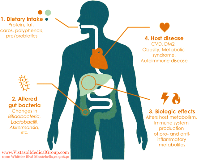
New research — using mouse models and fecal samples collected from humans — looks into the mechanisms that promote insulin resistance via the gut environment. The type of diet a person eats may be key, the researchers suggest.

New research looks in more detail at the mechanisms linking diet to insulin resistance.

Insulin resistance occurs when the body stops responding normally to insulin, a hormone that helps the body process sugar.

Developing insulin resistance can lead to type 2 diabetes, which is a metabolic condition that affects millions of people worldwide.

Obesity is a significant risk factor for insulin resistance and diabetes. But how and why does obesity drive this metabolic change?

Researchers from the University of Toronto in Canada believe the answer may lie in the mechanisms that consuming a high fat diet sets in motion.

“During high fat diet feeding and obesity, a significant shift occurs in the microbial populations within the gut, known as dysbiosis, which interacts with the intestinal immune system,” the researchers explain in their new study paper, published in Nature Communications.

The team decided to try and find out exactly how a high fat diet might alter gut immunity and, thus, bacterial balance, leading to insulin resistance.

“A link between the gut microbiota and the intestinal immune system is the immune derived molecule immunoglobulin A (IgA),” the researchers note in their paper. They add that this molecule is an antibody produced by B cells, a type of immune cells.

The investigators thought that IgA might be the missing link that explained how a poor diet leads to insulin resistance by altering gut immunity.

A sensitive mechanism impacted by diet

In the first part of their study, the investigators used mouse models with obesity, some of which lacked IgA. The researchers found that when the IgA-deficient mice ate a high fat diet, their insulin resistance worsened.

When the researchers collected gut bacteria from the IgA-deficient mice and transplanted them into rodents without gut bacteria, these mice also developed insulin resistance.

This experiment, the researchers suggest, indicates that at normal levels, IgA would help keep gut bacteria in check. Not just that, but it would also help prevent harmful bacteria from “leaking” through the intestines.

Mice without IgA had increased gut permeability, meaning that harmful bacteria could “leak” from the gut into the rest of the body.

Following these experiments in preclinical models, the researchers then moved on to see if the same mechanisms applied to humans. They were able to obtain stool samples from individuals who had undergone bariatric surgery — a form of surgery for weight loss.

The researchers analyzed the content of IgA in stool samples collected both before and after the individuals had undergone bariatric surgery.

The researchers found that these individuals had higher levels of IgA in their feces after surgery, suggesting that this antibody was indeed linked to metabolic function and influenced by diet.

“We discovered that during obesity, there are lower levels of a type of B cell in the gut that make an antibody called IgA,” notes the study’s lead author Helen Luck.

“IgA is naturally produced by our bodies and is crucial to regulating the bacteria that live in our gut,” she explains. She adds that “[i]t acts as a defense mechanism that helps neutralize potentially dangerous bacteria that take advantage of changes to the environment, such as when we consume an imbalanced or fatty diet.”

The results of the current research suggest a direct link between eating a high fat diet and having obesity, on the one hand, and having lower levels of gut IgA, symptoms of gut inflammation, and developing insulin resistance, on the other.

In the future, the researchers would like to find out how best to boost levels of IgA-producing B cells, believing that this intervention could protect against insulin resistance.

“If we can boost these IgA B cells or their products, then we may be able to control the type of bacteria in the gut. Especially the ones that are more likely to be linked to inflammation and ultimately, insulin resistance. “

Co-author Dr. Daniel Winer

“Going forward, this work could form the basis for new gut immune biomarkers or therapies for obesity and its complications, like insulin resistance and type 2 diabetes,” says study co-author Dr. Daniel Winer.

Both blood pressure numbers may predict heart disease


According to new research, both high systolic and high diastolic blood pressure can lead to heart attack and stroke.

Heart disease and stroke are the leading causes of death worldwide. In the United States, more than 600,000 people die of heart disease every year. According to the Centers for Disease Control and Prevention (CDC), nearly one-quarter of deaths due to cardiovascular disease are preventable.

Blood pressure readings are critical for analyzing and monitoring blood pressure. These tests record blood pressure using two measurements: systolic and diastolic blood pressure. Understanding these numbers is key to controlling blood pressure.

The systolic pressure shows how much pressure the blood places on the arteries when the heart beats, while the diastolic blood pressure shows the pressure while the heart is resting between beats. The American Heart Association (AHA) advise that blood pressure numbers below 120/80 millimeters of mercury (mm Hg) are normal.

When readings range from 120–129 mm Hg systolic and less than 80 mm Hg diastolic, the person has elevated blood pressure. Hypertension occurs when blood pressure is consistently over 130 mm Hg systolic or more than 80 mm Hg diastolic.

Which number is more important?

When doctors evaluate the risk of high blood pressure, they usually pay more attention to systolic blood pressure, which they consider a major risk factor for cardiovascular disease in older adults.

Decades of research have indicated that high systolic blood pressure is more likely than diastolic pressure to predict heart disease, but now, a new study finds that both numbers in blood pressure readings have a strong association with heart attack and stroke risk.

Researchers at Kaiser Permanente, a healthcare company in Oakland, CA, carried out the study, which appears in the New England Journal of Medicine.

The research involved more than 36 million blood pressure readings from 1.3 million people. The results challenged previous findings and showed the importance of both systolic and diastolic blood pressure.

“This research brings a large amount of data to bear on a basic question, and it gives such a clear answer,” says Kaiser Permanente stroke specialist Dr. Alexander C. Flint, who is the lead author of the study.

The study’s senior author is Dr. Deepak L. Bhatt, executive director of Interventional Cardiovascular Services at Brigham and Women’s Hospital and professor of medicine at Harvard Medical School — both in Boston, MA.

Dr. Flint explains that previous research has influenced cardiology guidelines, which have focused primarily on systolic pressure to predict the risk of heart disease. Some experts even argue that it might be possible to ignore the diastolic number.

The new study is the largest of its kind. The findings confirmed that systolic pressure has a greater effect, but they also demonstrated that both systolic and diastolic pressure can predict the risk of heart attack or stroke.

The researchers analyzed the effects of systolic and diastolic hypertension on a variety of adverse outcomes, such as “myocardial infarction, ischemic stroke, or hemorrhagic stroke,” over 8 years and found that both components independently predicted heart attack and stroke.

The recently updated American College of Cardiology and AHA guidelines now recommend more closely monitoring people at increased risk of high blood pressure. The findings of the new study that both systolic and diastolic hypertension have an effect at the lower threshold of 130/80 mm Hg support this change.

The National Institutes of Health’s Systolic Blood Pressure Intervention Trial (SPRINT) has also produced similar results.

“This analysis, using a very large amount of longitudinal data, convincingly demonstrates that both are important, and it shows that in people who are otherwise generally healthy, lower blood pressure numbers are better.”

Urine test can help diagnose aggressive prostate cancer

Recent research has revealed that a new urine test can detect aggressive prostate cancer cases that need treatment up to 5 years sooner than other diagnostic methods.

Researchers from the University of East Anglia (UEA) in Norwich, United Kingdom, and the Norfolk and Norwich University Hospital (NNUH) carried out the study.

They revealed that an experimental urine test, called Prostate Urine Risk (PUR), can find cancers that will require treatment within the first 5 years of diagnosis.

The findings now appear in the journal BJU International.

The team included Prof. Colin Cooper, Dr. Daniel Brewer, and Dr. Jeremy Clark, from UEA’s Norwich Medical School. Rob Mills, Marcel Hanna, and Prof. Richard Ball, of the NNUH, provided support.

Looking at biomarkers

To develop this unique test, the researchers looked at gene expression in the urine samples of 535 men and determined the cell-free expression of 167 different genes.

They then established a combination of 35 different genes that the scientists considered risk signatures, or biomarkers, that the PUR test could look for.

This test is unique in that it can sort people into different risk groups, thereby demonstrating the aggressiveness of the cancer.

“This research shows that our urine test could be used to not only diagnose prostate cancer without the need for an invasive needle biopsy but to identify a [person’s] level of risk,” says Dr. Clark.

“This means that we could predict whether or not prostate cancer patients already on active surveillance would require treatment. The really exciting thing is that the test predicted disease progression up to 5 years before it was detected by standard clinical methods.”

“Furthermore,” he adds, “the test was able to identify men that were up to eight times less likely to need treatment within 5 years of diagnosis.”

Prostate cancer is common but slow-growing

According to the American Cancer Society (ACS), around 1 in 9 men will receive a diagnosis of prostate cancer during their lifetime. In 2019, the ACS estimate that there will be around 174,000 new cases of prostate cancer and over 31,000 deaths from the condition.

That said, most cases of prostate cancer do not result in death. In fact, the 5-year survival rate for localized and regional prostate cancer is nearly 100%, and even when combined with those who have distant-stage prostate cancer, the overall survival rate is still 98%.

Not counting skin cancer, prostate cancer is the most common cancer among men. Thanks to early detection techniques, doctors can diagnose and treat many cases early. Because it is a slow growing cancer, tests usually find before it before it has the chance to spread.

What this test means in a clinical setting

There are many ways to help identify prostate cancer. Although a prostate biopsy is the only way to definitely diagnose the condition, there are a few screening tests that can indicate if a biopsy is necessary.

For example, the prostate-specific antigen (PSA) blood test can help detect the possible presence of prostate cancer. Doctors tend to use these results, or a series of results, to determine if someone needs a biopsy.

Doctors might also perform a digital rectal exam to see if there are areas on the prostate that could be cancer. Although it is less effective than a PSA test, it can sometimes find cancers in people with normal PSA levels.

The PUR test goes one step further; it not only identifies the presence of cancer earlier than other tests, it can also help put people into different risk groups so that doctors can more accurately determine the course of care and whether to watch and wait, take a biopsy, or start treatment immediately.

“If this test was to be used in the clinic, large numbers of men could avoid an unnecessary initial biopsy and the repeated, invasive follow-up of men with low-risk disease could be drastically reduced.”

When to see a doctor if a child has a nosebleed

Nosebleeds are common in children but are usually short-lived and rarely a cause for concern. Persistent, recurring, or very heavy bleeding may, however, require medical attention.

Doctors refer to nosebleeds as epistaxis. Approximately 60 percent of people will experience a nosebleed at some point during their life. However, nosebleeds occur most commonly in children aged between 2 and 10 years and in older people aged 50 to 80 years.

Although the bleeding can sometimes be alarming, only about 10 percent of nosebleeds are serious enough to require medical treatment.

In this article, we explain what to do when a child’s nose starts bleeding and when to see a doctor. We also discuss medical treatments, causes, and tips for prevention.

What to do

A person can usually treat a child’s nosebleed at home. It is important to stay calm because most nosebleeds are short-lived and do not indicate a serious problem.

To treat a child with a nosebleed:

  • Start by sitting the child down and reassuring them. Have them sit upright and leaning slightly forward.
  • Do not lean the child back or lie them down because this can cause them to swallow the blood and may lead to coughing or vomiting.
  • Gently pinch the tip of the child’s nose between two fingers using a tissue or clean towel and have them breathe through their mouth.
  • Continue to apply pressure for around 10 minutes, even if the bleeding stops.

Do not fill the child’s nose with gauze or tissue and avoid spraying anything into the nose.

When to see a doctor

Children with nosebleeds do not typically require medical attention. Most nosebleeds are short-lived, and it is usually possible to treat the child at home.

However, talk to a doctor if the nosebleeds:

  • occur frequently
  • change from a familiar pattern to a new one
  • occur alongside chronic congestion or other signs of easy bleeding or bruising
  • begin after the child starts taking a new medication
  • regularly require a trip to the emergency room

A nosebleed requires urgent medical attention if:

  • it continues after 20 minutes of applying pressure to the child’s nose
  • it occurs following a head injury, fall, or blow to the face
  • the child also has an intense headache, a fever, or other concerning symptoms
  • the child’s nose appears misshapen or broken
  • the child shows signs of having lost too much blood, such as looking pale, having little energy, feeling dizzy, or passing out
  • the child begins coughing up or vomiting blood
  • the child has a bleeding disorder or is taking blood thinners

Medical treatment

Children with severe nosebleeds should see a healthcare professional, who will try to stop the bleeding.

Treatment options for nosebleeds include:

  • applying silver nitrate to blood vessels to seal them
  • cauterizing, or burning, the blood vessels to seal them
  • packing the nose with medicated gauze to constrict the blood vessels

After stopping the bleeding, a doctor will examine the child to determine the cause. In some cases, the child may require surgery to fix a problem with the blood vessels in the nose.

Causes

An injury or blow to the face can irritate blood vessels in the nose.

Most nosebleeds are anterior nosebleeds, which means that the bleeding occurs in the front, soft part of the nose. This area of the nose contains many small blood vessels that can rupture and bleed if they become irritated or inflamed.

Posterior nosebleeds develop in the rear of the nose and rarely occur in children. This type of nosebleed tends to be heavier, and it can be more difficult to stop the bleeding.

Irritation of the blood vessels is a common cause of anterior nosebleeds. Several things can irritate the blood vessels in the nose, including:

  • dry air
  • picking the nose
  • nasal allergies
  • an injury or blow to the nose or face, for example, from a ball or fall
  • sinusitis, common colds, the flu, and other infections that affect the nasal passages
  • nasal polyps
  • overuse of nasal sprays

Less common causes of nosebleeds in children can include:

  • conditions that affect bleeding or blood clotting, such as hemophilia
  • certain medications, including blood thinners
  • heart disease
  • high blood pressure
  • cancer

Prevention tips

Although it may not be possible to prevent all nosebleeds in children, a person can take steps to help reduce their occurrence. These include:

  • treating allergies to prevent inflammation in the nose
  • using saline (saltwater) nasal sprays to keep the child’s nose moist
  • running a humidifier or vaporizer in the child’s bedroom to prevent the air from drying out
  • keeping children’s nails trimmed to prevent injuries due to nose picking
  • encouraging children to wear appropriate protective equipment during sports or other activities where injury to the nose is possible

Summary

Nosebleeds are a common occurrence in young children and rarely a cause for concern. A person can usually treat the bleeding at home by applying continuous, gentle pressure to the soft part of the child’s nose for around 10 minutes.

Call 911 or take the child to the emergency room if they seem dizzy or weak or if they pass out. It is also necessary to seek immediate medical attention if the bleeding is very heavy, does not stop after 20 minutes, or occurs after a fall or head injury.

Most nosebleeds in children are due to dry air, nose picking, nasal allergies, or other factors that irritate the delicate blood vessels in the front of the nose.

A person should consult a doctor or pediatrician if the child has frequent nosebleeds or has recently started taking a new medication.

What is obesity and what it causes?

Calories Sedentary lifestyle Not sleeping enough Endocrine disruptors Medications Is obesity self-perpetuating? Obesity gene Takeaway

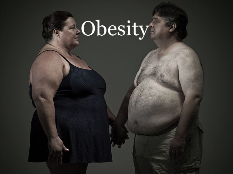
Obesity is a medical condition that occurs when a person carries excess weight or body fat that might affect their health. A doctor will usually suggest that a person has obesity if they have a high body mass index.

Body mass index (BMI) is a tool that doctors use to assess if a person is at an appropriate weight for their age, sex, and height. The measurement combines height and weight.

A BMI between 25 and 29.9 indicates that a person is carrying excess weight. A BMI of 30 or over suggests that a person may have obesity.

Other factors, such as the ratio of waist-to-hip size (WHR), waist-to-height ratio (WtHR), and the amount and distribution of fat on the body also play a role in determining how healthy a person’s weight and body shape are.

If a person does have obesity and excess weight, this can increase their risk of developing a number of health conditions, including metabolic syndrome, arthritis, and some types of cancer.

Metabolic syndrome involves a collection of issues, such as high blood pressure, type 2 diabetes, and cardiovascular disease.

Maintaining a healthy weight or losing through diet and exercise is one way to prevent or reduce obesity. In some cases, a person may need surgery.

Now read on to find out why obesity happens.

1) Consuming too many calories

When a person consumes more calories than they use as energy, their body will store the extra calories as fat. This can lead to excess weight and obesity.

Also, some types of foods are more likely to lead to weight gain, especially those that are high in fats and sugars.

Foods that tend to increase the risk of weight gain include:

  • fast foods
  • fried foods, such as french fries
  • fatty and processed meats
  • many dairy products
  • foods with added sugar, such as baked goods, ready-made breakfast cereals, and cookies
  • foods containing hidden sugars, such as ketchup and many other canned and packaged food items
  • sweetened juices, sodas, and alcoholic drinks
  • processed, high-carb foods, such as bread and bagels

Some processed food products contain high-fructose corn syrup as a sweetener, including savory items, such as ketchup.

Eating too much of these foods and doing too little exercise can result in weight gain and obesity.

A person who consumes a diet that consists mainly of fruits, vegetables, whole grains, and water is still at risk of gaining excess weight if they overeat, or if genetic factors, for example, increase their risk.

However, they are more likely to enjoy a varied diet while maintaining a healthy weight. Fresh foods and whole grains contain fiber, which makes a person feel full for longer and encourages healthy digestion.

2) Leading a sedentary lifestyle

Many people lead a much more sedentary lifestyle than their parents and grandparents did.

Examples of sedentary habits include:

  • working in an office rather than doing manual labor
  • playing games on a computer instead of doing physical activities outside
  • going to places by car instead of walking or cycling

The less a person moves around, the fewer calories they burn.

Also, physical activity affects how a person’s hormones work, and hormones have an impact on how the body processes food.

Several studies have shown that physical activity can help to keep insulin levels stable and that unstable insulin levels may lead to weight gain.

Researchers who published a review in BMJ Open Sport and Exercise Medicine in 2017 noted that, while the designs of some studies make it hard to draw exact conclusions, “A lifestyle incorporating regular [physical activity] has been identified as a key factor for maintaining and improving many aspects of health, including insulin sensitivity.”

Physical activity need not be training in the gym. Physical work, walking or cycling, climbing stairs, and household tasks all contribute.

However, the type and intensity of activity may affect the degree to which it benefits the body in the short- and long-term.

3) Not sleeping enough

Research has suggested that missing sleep increases the risk of gaining weight and developing obesity.

Researchers reviewed evidence for over 28,000 children and 15,000 adults in the United Kingdom from 1977 to 2012. In 2012, they concluded that sleep deprivation significantly increased obesity risk in both adults and children.

The changes affected children as young as 5 years of age.

The team suggested that sleep deprivation may lead to obesity because it can lead to hormonal changes that increase the appetite.

When a person does not sleep enough, their body produces ghrelin, a hormone that stimulates appetite. At the same time, a lack of sleep also results in a lower production of leptin, a hormone that suppresses the appetite.

4) Endocrine disruptors

A team from the University of Barcelona published a study in the World Journal of Gastroenterology that provides clues as to how liquid fructose — a type of sugar — in beverages may alter lipid energy metabolism and lead to fatty liver and metabolic syndrome.

Features of metabolic syndrome include diabetes, cardiovascular disease, and high blood pressure. People with obesity are more likely to have metabolic syndrome.

After feeding rats a 10-percent fructose solution for 14 days, the scientists noted that their metabolism was starting to change.

Scientists believe there is a link between high consumption of fructose and obesity and metabolic syndrome. Authorities have raised concerns about the use of high-fructose corn syrup to sweeten drinks and other food products.

Animal studies have found that when obesity occurs due to fructose consumption, there is also a close link with type 2 diabetes.

In 2018, researchers published the results of investigations involving young rats. They, too experienced metabolic changes, oxidative stress, and inflammation after consuming fructose syrup.

The researchers note that “increased fructose intake may be an important predictor of metabolic risk in young people.”

They call for changes in the diets of young people to prevent these problems.

Avoiding high-fructose corn syrup

Foods that contain high-fructose corn syrup include:

  • sodas, energy drinks, and sports drinks
  • candy and ice cream
  • coffee creamer
  • sauces and condiments, including salad dressings, ketchup, and barbecue sauce
  • sweetened foods, such as yogurt, juices, and canned foods
  • bread and other ready-made baked goods
  • breakfast cereal, cereal bars, and “energy” or “nutrition” bars

To reduce your intake of corn syrup and other additives:

  • check the labels before you buy
  • opt for unsweetened or less processed items where possible
  • make salad dressings and bake other products at home

Some foods contain other sweeteners, but these can also have adverse effects.

5) Medications and weight gain

Some medications can also lead to weight gain.

Results of a review and meta-analysis published in The Journal of Clinical Endocrinology and Metabolism in 2015 found that some medicines caused people to gain weight over a period of months.

  • atypical antipsychotics, especially olanzapine, quetiapine, and risperidone
  • anticonvulsants and mood stabilizers, and specifically gabapentin
  • hypoglycemic medications, such as tolbutamide
  • glucocorticoids used to treat rheumatoid arthritis
  • some antidepressants

However, some medications may lead to weight loss. Anyone who is starting a new medication and is concerned about their weight should ask their doctor whether the drug is likely to have any effect on weight.

6) Is obesity self-perpetuating?

The longer a person is overweight, the harder it may be for them to lose weight.

Findings of a mouse study, published in the journal Nature Communications in 2015, suggested that the more fat a person carries, the less likely the body is to burn fat, because of a protein, or gene, known as sLR11.

It seems that the more fat a person has, the more sLR11 their body will produce. The protein blocks the body’s ability to burn fat, making it harder to shed the extra weight.

7) Obesity gene

A faulty gene called the fat-mass and obesity-associated gene (FTO) is responsible for some cases of obesity.

A study published in 2013 points to a link between this gene and:

  • obesity
  • behaviors that lead to obesity
  • a higher food intake
  • a preference for high-calorie foods
  • an impaired ability to feel full, known as satiety

The hormone ghrelin plays a crucial role in eating behavior. Ghrelin also affects the release of growth hormones and how the body accumulates fat, among other functions.

The activity of the FTO gene might impact a person’s chances of having obesity because it affects the amounts of ghrelin a person has.

In a study involving 250 people with eating disorders, published in Plos One in 2017, researchers suggested that aspects of FTO might also play a role in conditions, such as binge eating and emotional eating.

Takeaway

Many factors play a role in the development of obesity. Genetic traits can increase the risk in some people.

A healthful diet that contains plenty of fresh food, together with regular exercise, will reduce the risk of obesity in most people.

However, those that have a genetic predisposition may find it harder to maintain a healthy weight.